A) Preoperative intraoral periapical (IOPA) radiograph of 36. B) Post operative (IOPA) radiograph of 36. C) 1 month follow up IOPA radiograph of 36. D) 6 months follow up IOPA radiograph of

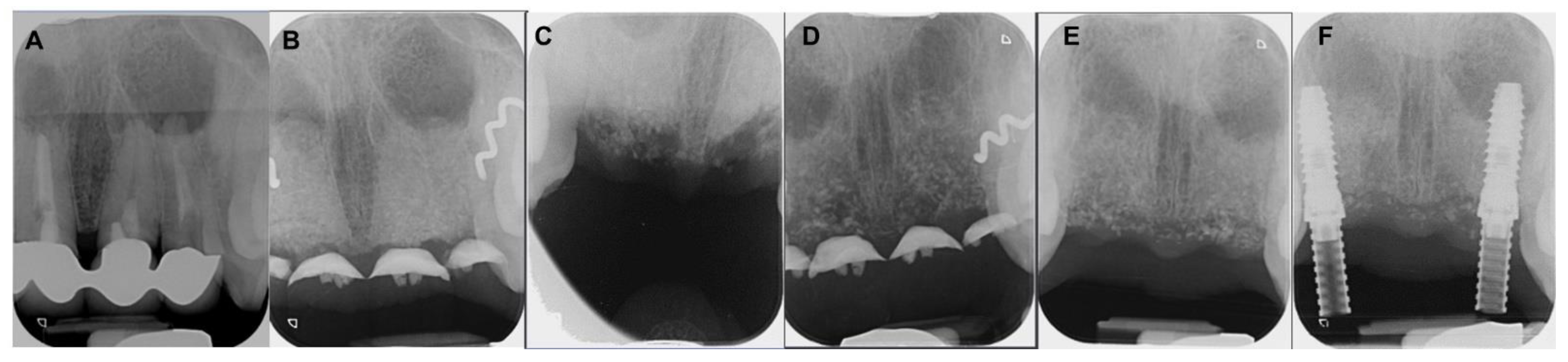
A) Preoperative intraoral periapical (IOPA) radiograph of 36. B) Post operative (IOPA) radiograph of 36. C) 1 month follow up IOPA radiograph of 36. D) 6 months follow up IOPA radiograph of 36. E) 1 year follow up IOPA radiograph of 36. - IP Indian J Conserv Endod - clinical and preclinical conservative /restorative de
jcdr-14-ZD01-g003.jpg
Coatings, Free Full-Text
Effectiveness of Platelet Rich Plasma and Bone Graft in the Treatment of Intrabony Defects: A Clinico-radiographic Study

A) Preoperative intraoral periapical (IOPA) radiograph of 36. B) Post

Treatment of Deeply Carious Vital Primary Molars by Complete

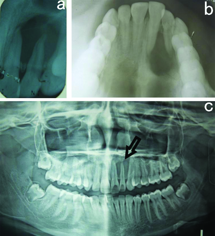
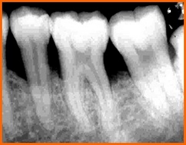
Preoperative radiograph of tooth 36 in conventional right angle
The advantages of pre-operative radiograph in the diagnosis and in

Postoperative IOPA of mandibular left side depicting no bone gain

Radiograph sem