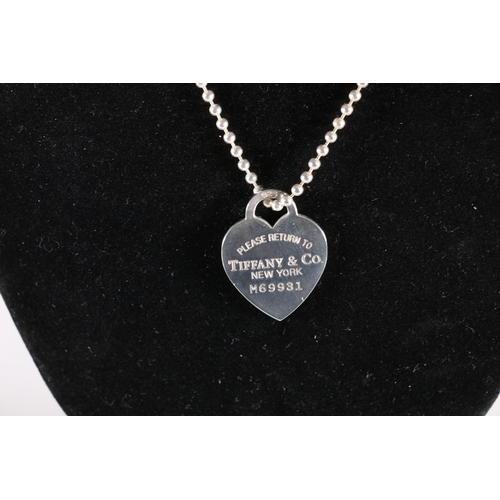
Anatomy, descriptive and applied. Anatomy. 556 THE VASCULAR SYSTEMS The anterior cardiac veins open into the lower fore part of the right auricle. The right auriculoventricular opening, or the tricuspid orifice {
Download this stock image: . Anatomy, descriptive and applied. Anatomy. 556 THE VASCULAR SYSTEMS The anterior cardiac veins open into the lower fore part of the right auricle. The right auriculoventricular opening, or the tricuspid orifice {ostium venosum dexirum), is the large oval aperture of communication between the right auricle and the ventricle; it will be described with the right ventricle. The Eustachian valve (valvula venae cavae inferioris [Eustachii]) is situated in front of the orifice of the inferior vena cava. It is semilunar in form, its convex margin being attached to the anterior margin of the inferior - RN5967 from Alamy's library of millions of high resolution stock photos, illustrations and vectors.

What is the real cardiac anatomy, clinical anatomy 2019

JaypeeDigital
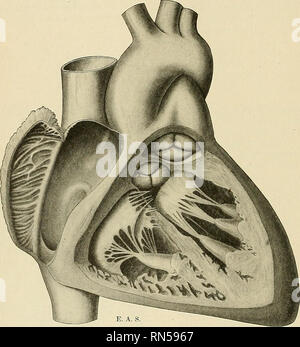
Right-Sided Obstructive Lesions (Section 2) - Congenital Cardiac Anesthesia

Clinical Anatomy (A Problem Solving Approach), 2nd Edition PDF, PDF, Common Carotid Artery

Tricuspid valve hi-res stock photography and images - Page 5 - Alamy

Eustachii hi-res stock photography and images - Alamy

Anatomy, descriptive and applied. Anatomy. CARDIAC VEINS 709 1. The great cardiac or left coronary vein {v. cordis magna) begins at the apex of the heart and ascends along the anterior

JaypeeDigital

Organs of the Cardiovascular System and their Neurovasculature
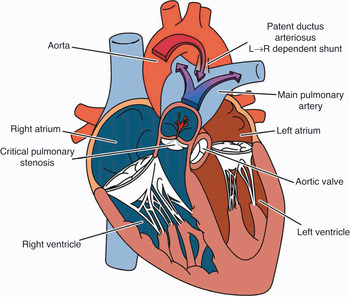
Coronary artery anomalies overview: The normal and the abnormal