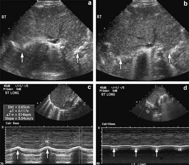
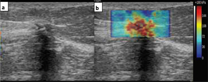
Representative ultrasound B-mode and elastography images for (A)

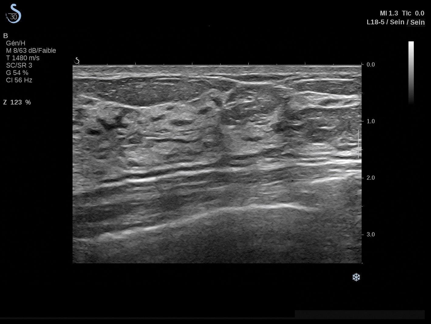
a) The gray-scale image of mass
/guideline/2022/img/t/k_cq4_t2.png

Comparison of portal vein hemodynamics with ultrasound-based elastography for the prediction of liver fibrosis in patients with chronic liver disease

Representative B-mode and elastography images acquired during and after

Takanori WATANABE, National Hospital Organization Sendai Medical Center, Sendai, breast surgery

PDF) Color Doppler to Characterize Malignant Breast Lesion

PDF) Breast Elastography: A Hospital-Based Preliminary Study in China

B-mode ultrasound, color Doppler, and sonoelastography in differentiation between benign and malignant cervical lymph nodes with special emphasis on sonoelastography, Egyptian Journal of Radiology and Nuclear Medicine
Diagnostic utility of strain and shear wave ultrasound elastography in differentiation of benign and malignant solid breast lesions, Egyptian Journal of Radiology and Nuclear Medicine

PDF] Ultrasound elastography improves differentiation between benign and malignant breast lumps using B-mode ultrasound and color Doppler

Ultrasound elastography in peripheral cervical lymph nodes. A Color