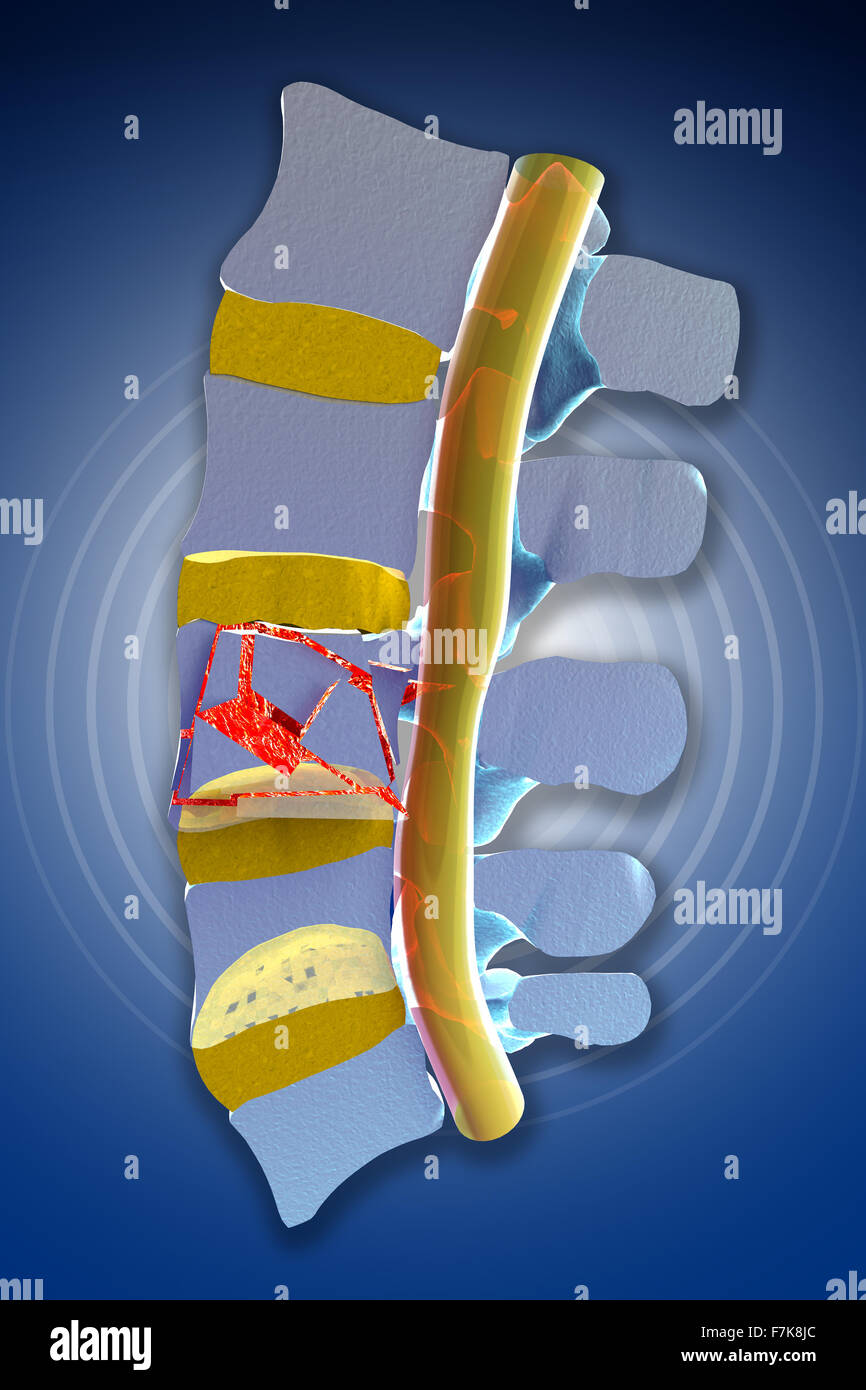
Lumbar Compression Fracture, Illustration - Stock Image - C027

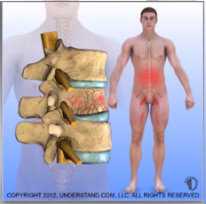
An interpretive illustration of an MRI depicting a sagittal view of compression fractures at the L1 and L2 vertebrae as a result of osteoporosis. Over time as bone becomes weaker and more porous, they become more susceptible to injury and fractures, especially in situations where significant weight or stress is placed on the bone. Evan Oto/SCIENCE PHOTO LIBRARY

Lumbar Compression Fracture, MRI - Stock Image - C027/1372

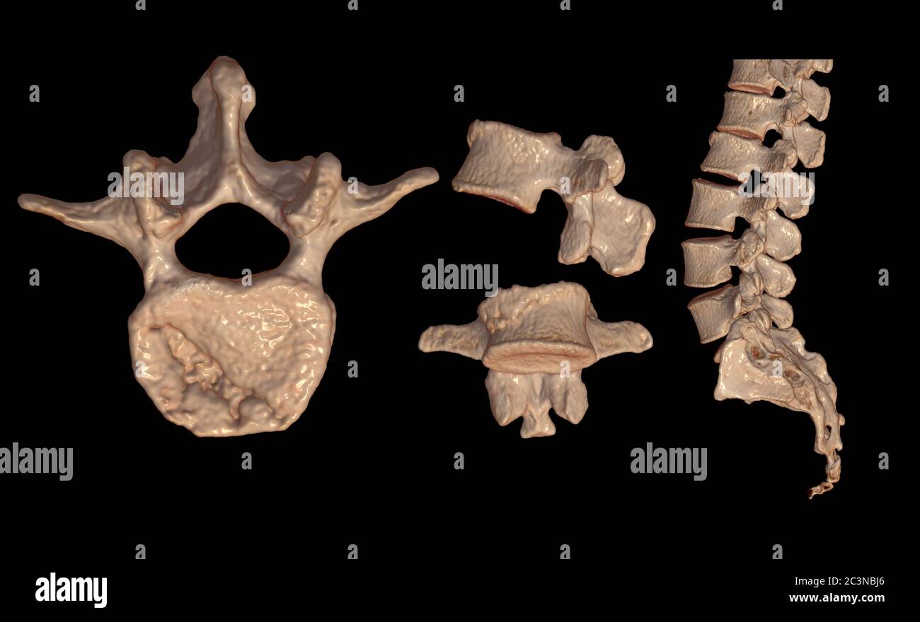
Thoracic Vertebrae Compression Fracture - Stock Image - C027/1223

999 Compression Fracture Stock Photos - Free & Royalty-Free Stock
110+ Compression Fracture Spine Stock Illustrations, Royalty-Free

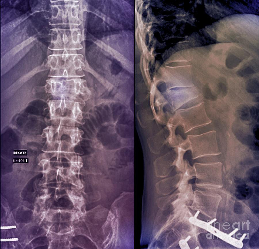
Thoracic vertebral collapse, X-ray - Stock Image - C038/6661

x ray of compression fracture - Keyword Search - Science Photo Library

Free Vectors Two types of compression fractures of the lumbar
Compression fractures hi-res stock photography and images - Alamy

Lumbar Vertebral Body Compression Fractures - Stock Image - C043
Compression Fracture Of A Lumbar Vertebra #2 Photograph by Zephyr
Compression fracture spine hi-res stock photography and images - Alamy

127 Vertebral Compression Fracture Stock Photos - Free & Royalty

103 Spine Compression Fracture Stock Photos, High-Res Pictures

127 Vertebral Compression Fracture Stock Photos - Free & Royalty
Virginia Interventional Pain & Spine Centers