Finite element analysis of compression fractures at the thoracolumbar junction using models constructed from medical images
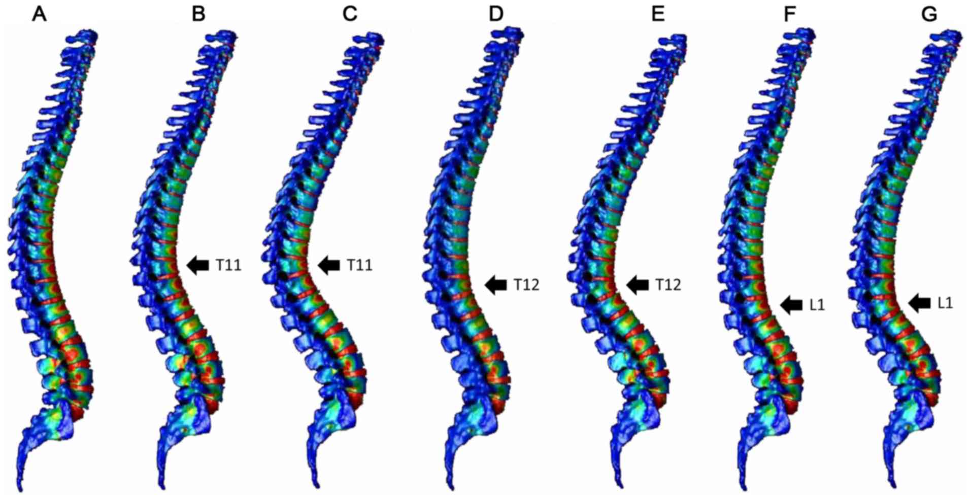
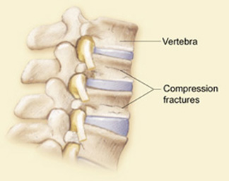
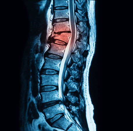
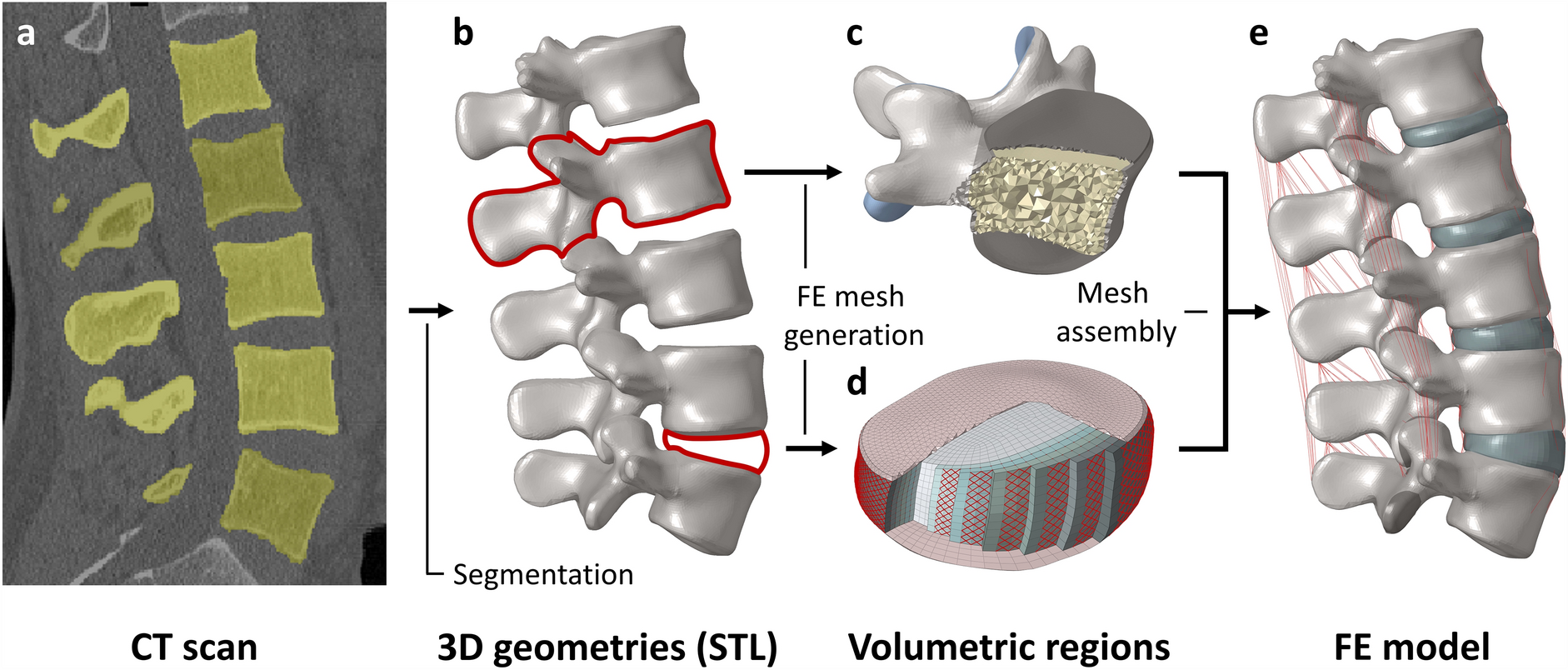
Vertebral fractures commonly occur at the thoracolumbar junction. These fractures can be treated with mild residual deformity in many cases, but are reportedly associated with increased risk of secondary vertebral fractures. In the present study, a three‑dimensional (3D) whole spine model was constructed using the finite element method to explore the mechanism of development of compression fractures. The 3D model of the whole spine, from the cervical spine to the pelvis, was constructed from computed tomography (CT) images of an adult male. Using a normal spine model and spine models with compression fractures at the T11, T12 or L1 vertebrae, the distribution of strain was analyzed in the vertebrae after load application. The normal spine model demonstrated greater strain around the thoracolumbar junction and the middle thoracic spine, while the compression fracture models indicated focused strain at the fracture site and adjacent vertebrae. Increased load time resulted in the extension of the strain region up to the middle thoracic spine. The present findings, that secondary vertebral fractures commonly occur around the fracture site, and may also affect the thoracic vertebrae, are consistent with previous clinical and experimental results. These results suggest that follow‑up examinations of compression fractures at the thoracolumbar junction should include the thoracic spine and adjacent vertebrae. The current data also demonstrate that models created from CT images can be used for various analyses.

Bioengineering, Free Full-Text

JCM, Free Full-Text

Establishment and validation of a T12-L2 3D finite element model for thoracolumbar segments. - Abstract - Europe PMC

PDF] Finite element analysis of compression fractures at the thoracolumbar junction using models constructed from medical images

Vertebral compression fractures in the L1 vertebrae with TLF injury. A

Differences in surgical outcome after anterior corpectomy and reconstruction with an expandable cage with rectangular footplates between thoracolumbar and lumbar osteoporotic vertebral fracture - North American Spine Society Journal (NASSJ)
Comparison of anterior column reconstruction techniques after en bloc spondylectomy: a finite element study

Smoothed particle hydrodynamics implementation to enhance vertebral fracture finite element model in a cervical spine segment under compression - ScienceDirect

Compression fracture model construction. (a) Normal vertebral body. (b

Thoracolumbar flexion dysfunction and thoracolumbar compression fracture in postmenopausal women: a single-center retrospective study, Journal of Orthopaedic Surgery and Research

Applied Sciences, Free Full-Text