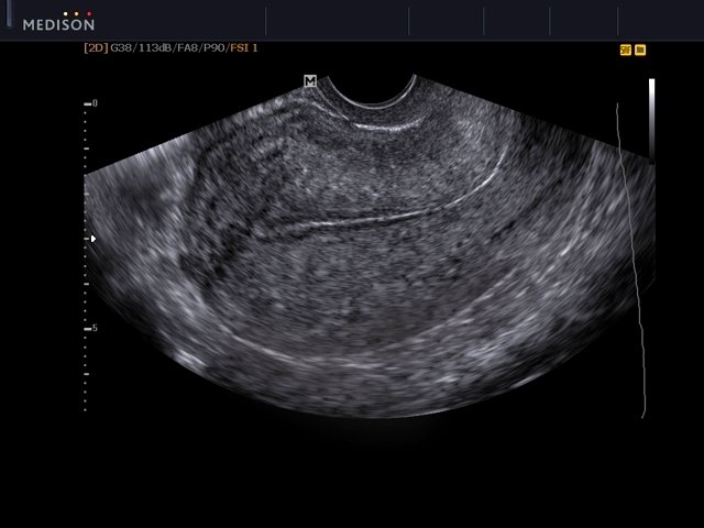
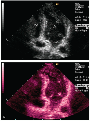
Download scientific diagram | Ultrasound imaging in B-mode, color and spectral Doppler of the abdominal organs of the agouti. (a) Ultrasonographic aspects of the urinary vesicle. Note the smooth and echogenic walls with a slight amount of sediment on the interior. (b,d) Color flow and B-mode renal morphology of the right and left kidneys, respectively, showing the usual echotexture and parenchymal echogenicity and preserved corticomedullary limit. (c,e) Pattern of flow of the renal artery, arcuate (arrowhead) and interlobar (arrow) arteries observed with color Doppler. The pulsed Doppler demonstrates well-defined systolic and diastolic peaks. from publication: Abdominal B-mode and Doppler ultrasonography of chemically restrained agouti (Dasyprocta prymnolopha Wagler, 1831) | Agoutis are small-sized wild animals whose body weight can reach up to 4kg, and are found throughout Brazil. They are considered important seed dispersers, especially for big trees and there are species that rely almost exclusively on these animals for their territorial | Doppler Ultrasonography, Doppler Ultrasound and Hemodynamics | ResearchGate, the professional network for scientists.

Pigmentation of spleens on subsequent weeks of life, and the

Applied Sciences, Free Full-Text

Determination of a potential quantitative measure of the state of the lung using lung ultrasound spectroscopy

Ultrasound imaging in B-mode, color and spectral Doppler of the
The Normande A br allele. (A) Genetic map representing the

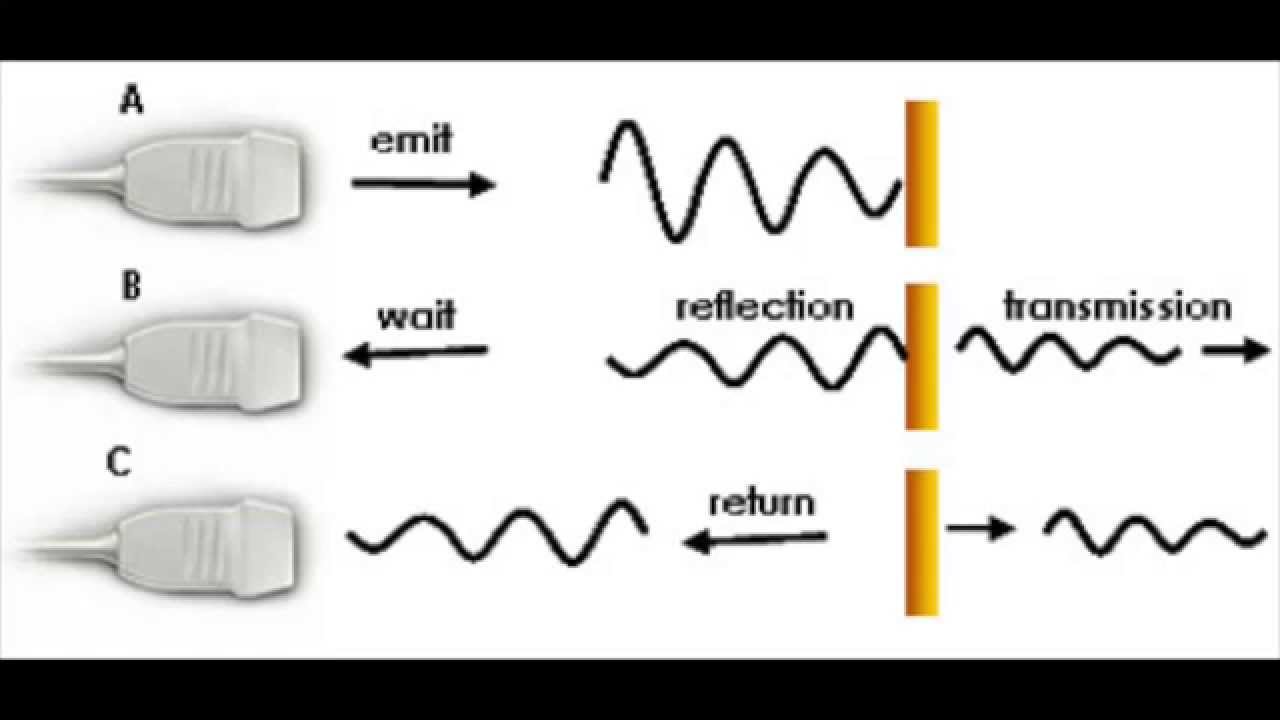
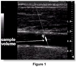
Example of a Doppler Spectrum with B-mode image for positioning.

Life, Free Full-Text

Ultrasound SpringerLink

A miniaturized ultrasound transducer for monitoring full-mouth oral health: a preliminary study
Specialized Echocardiographic Techniques and Methods

PDF) Abdominal B-mode and Doppler ultrasonography of chemically restrained agouti (Dasyprocta prymnolopha Wagler, 1831)
doppler applications