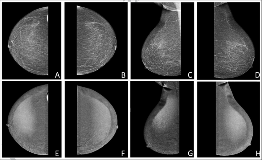
Download scientific diagram | Pre-contrast (A, B, C, D) and post-contrast recombined images (E, F, G, H) of both breasts demonstrating normal breast parenchyma with no suspicious findings. from publication: An initial experience of using dual energy contrast enhanced mammography at a tertiary care hospital in Pakistan | Objective Contrast enhanced mammography (CEM), a relatively new and promising modality, combines mammography (MMG) with an iodinated contrast material to illuminate neovascularity within the breast; analogous to magnetic resonance imaging (MRI). CEM improves the overall | Mammography, breast and Breast Cancer | ResearchGate, the professional network for scientists.
Metastasis of serous ovarian carcinoma to the breast: a case report and review of the literature, Journal of Medical Case Reports

Pre and postcontrast enhanced T1W MRI images (A and B) of the left

Pre-contrast (A&B) and post-contrast recombined images (C&D) of left
Contrast Media - Radiology at St. Vincent's University Hospital
Pre‐contrast (a) and post‐contrast (b) computed tomographic transverse

Post-contrast 3D T1-weighted TSE MR sequences (SPACE, CUBE, VISTA/BRAINVIEW, isoFSE, 3D MVOX): Technical aspects and clinical applications - ScienceDirect

Sana ZEESHAN, Assistant Professor

Radiographic contrast, Radiology Reference Article

PDF) An initial experience of using dual energy contrast enhanced