Commonly referred to as B (brightness) mode, the use of grey scale imaging in ultrasound renders a two-dimensional image in which the organs and tissues of interest are depicted as points of v
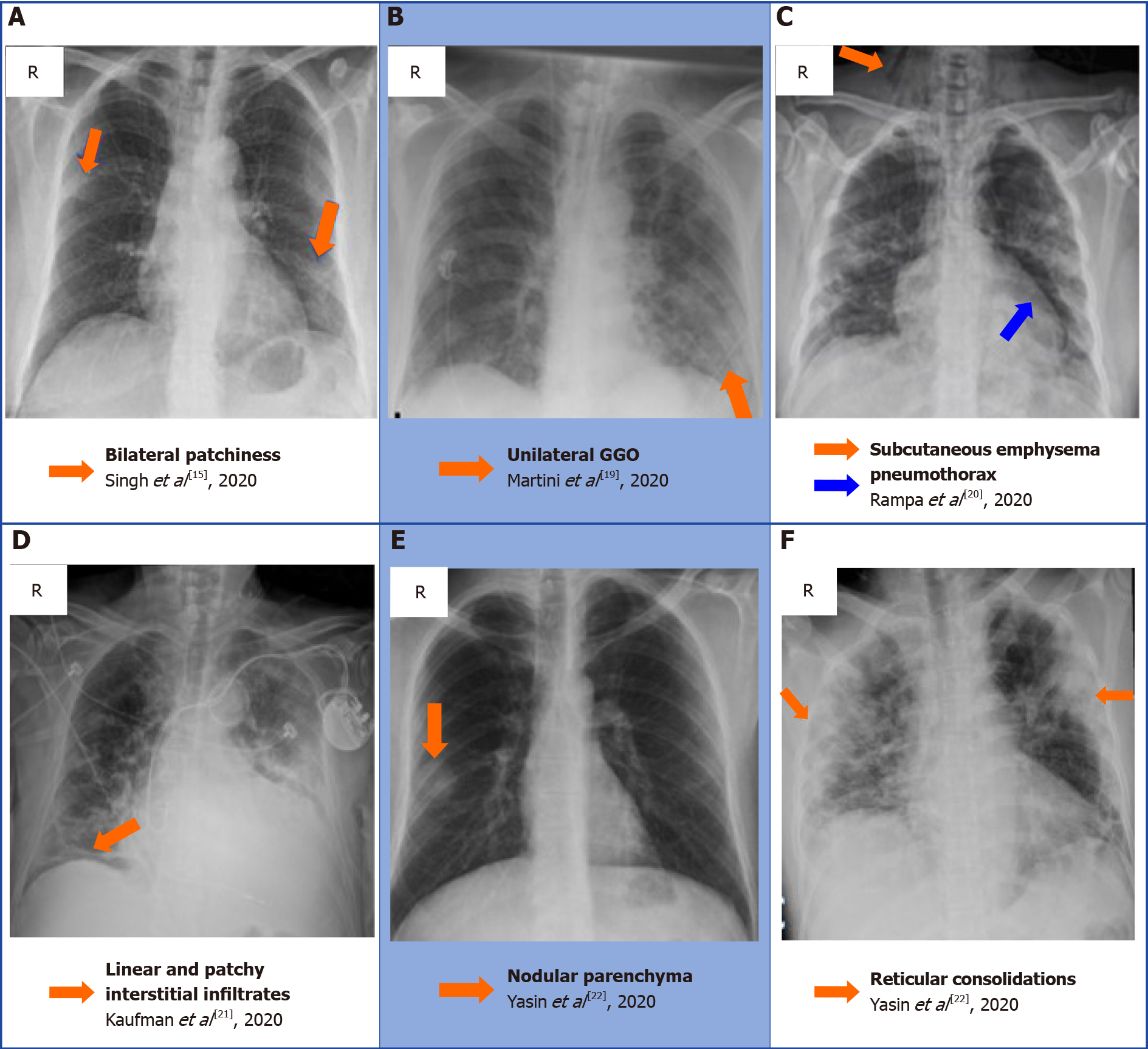
Comprehensive literature review on the radiographic findings, imaging modalities, and the role of radiology in the COVID-19 pandemic

Ultrasound grayscale ratio: a reliable parameter for differentiating between papillary thyroid microcarcinoma and micronodular goiter, BMC Endocrine Disorders

PDF] Muscle Ultrasound in Inflammatory Myopathies: A Critical Review

Development of an Ultrasound Scoring System to Describe Brain Maturation in Preterm Infants

Partial epilepsy: A pictorial review of 3 TESLA magnetic resonance imaging features – ScienceOpen
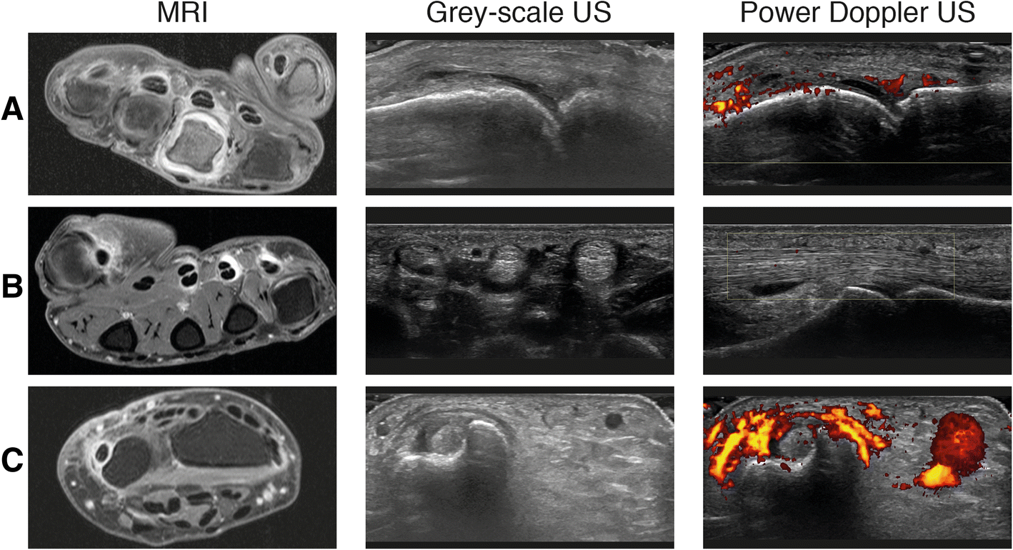
Do musculoskeletal ultrasound and magnetic resonance imaging identify synovitis and tenosynovitis at the same joints and tendons? A comparative study in early inflammatory arthritis and clinically suspect arthralgia

Stapes, Radiology Reference Article, stapes

Grayscale Ultrasound Artifacts

To explore the pathogenesis of Bell's palsy using diffusion tensor image

Image data of CBTs were collected on gray-scale, CDU, CTA, and MRA. (a)
Grey scale imaging (ultrasound), Radiology Reference Article
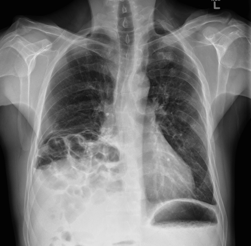
Diaphragmatic hernia - Radiology at St. Vincent's University Hospital
Grey-scale and Doppler ultrasound. Transverse (a) and sagittal (b) grey