Ultra-wide-field fundus photographs and ultra-wide-field

Download scientific diagram | Ultra-wide-field fundus photographs and ultra-wide-field fluorescein angiographic imaging of ocular toxocariasis. (A) A granuloma with mild vitreous opacity. (B) A tractional retinal fold with localized tractional retinal detachment. (C) Diffuse peripheral vascular leakage. (D) A prominent optic disc leakage. from publication: The Clinical Characteristics of Ocular Toxocariasis in Jeju Island Using Ultra-wide-field Fundus Photography | Toxocariasis, Ocular and Photography | ResearchGate, the professional network for scientists.
Ultra-widefield Imaging Ideal for Monitoring Myopic Maculopathy

Fundus photos of the patients for each case. (A) Case 1. Fundus image
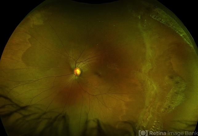
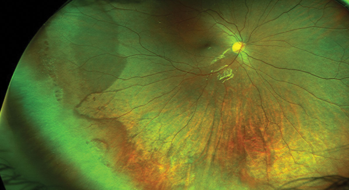
Ultra-Wide Field Fundus Photography Showing Lattice Degeneration - Retina Image Bank

Ultra-wide-field fundus photographs and ultra-wide-field fluorescein

Ultra-Widefield Fundus Photography Brisbane Eye Doctor Clinic & Ophthalmologist
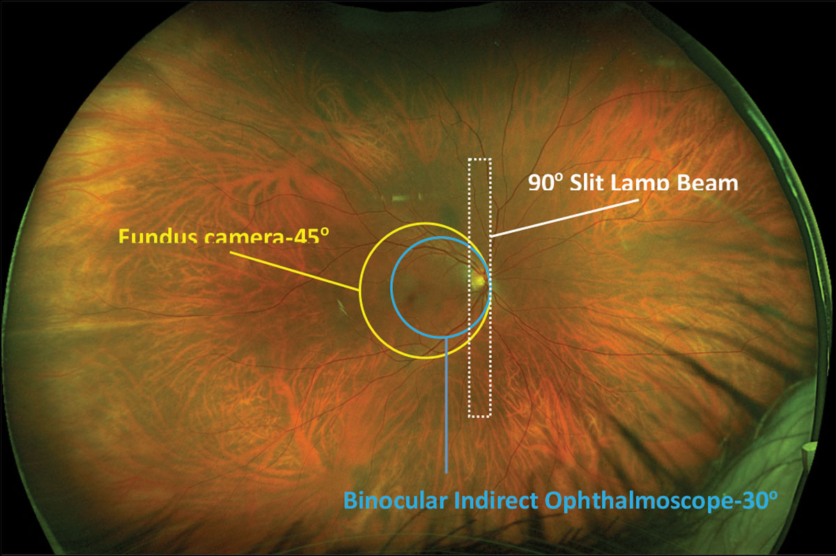
Ultra-Widefield Imaging: Expand Your Horizons
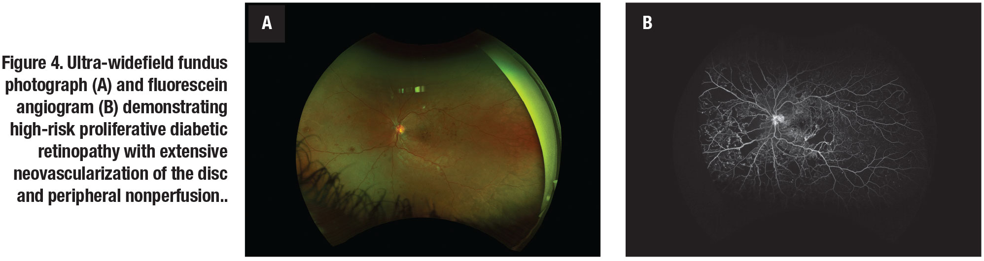
How ultra-widefield imaging is changing our view of DR

PDF) The Clinical Characteristics of Ocular Toxocariasis in Jeju Island Using Ultra-wide-field Fundus Photography
Ultra-Wide Field Retinal Imaging Device, Product Technology